File:EB1911 - Vascular System - Fig. 3.png

Size of this preview: 689 × 599 pixels. Other resolutions: 276 × 240 pixels | 552 × 480 pixels | 883 × 768 pixels | 1,029 × 895 pixels.
Original file (1,029 × 895 pixels, file size: 325 KB, MIME type: image/png)
| This is a file from the Wikimedia Commons. Information from its description page there is shown below. Commons is a freely licensed media file repository. You can help. |
Summary
| Description |
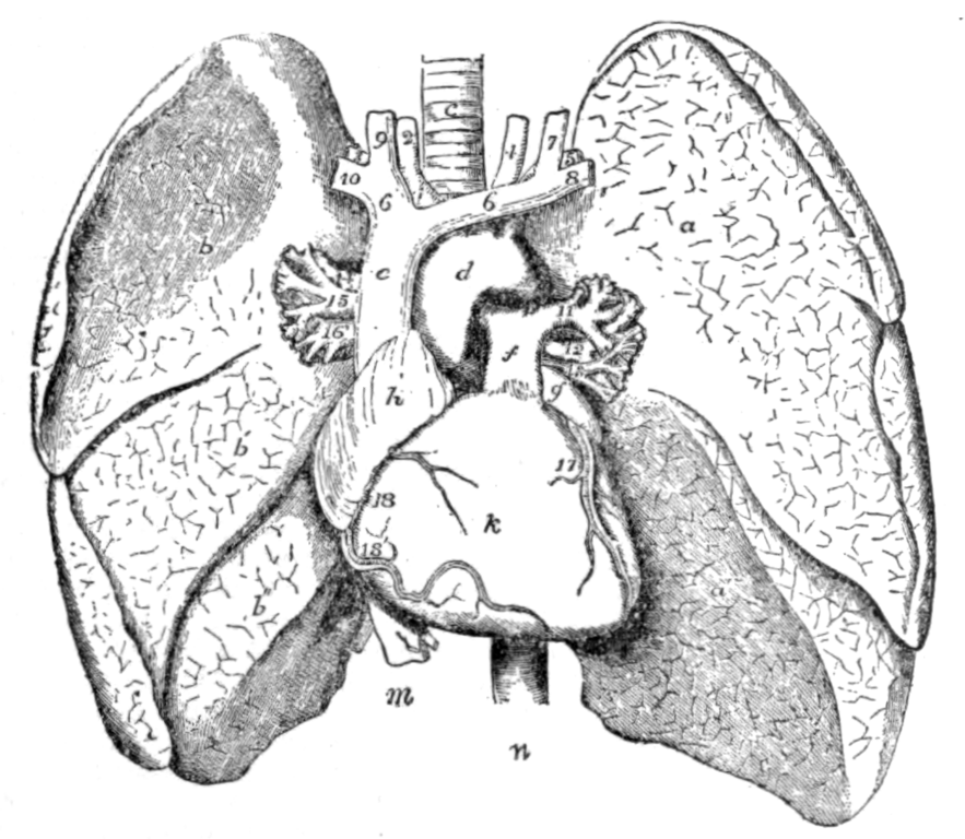
English: Fig. 3.—The Thoracic Viscera. In this diagram the lungs are turned to the side, and the pericardium removed to display the heart. a, upper, a′, lower lobe of left lung; b, upper, b′, middle, b″, lower lobe of right lung; c, trachea; d, arch of aorta; e, superior vena cava; f, pulmonary artery; g, left, and h, right auricle; k, right, and l, left ventricle; m, inferior vena cava; n, descending aorta; 1, innominate artery; 2, right, and 4, left common carotid artery; 3, right, and 5, left subclavian artery; 6, 6, right and left innominate vein; 7 and 9, left and right internal jugular veins; 8 and 10, left and right subclavian veins; 11, 12, 13, left pulmonary artery, bronchus and vein; 14, 15, 16, right pulmonary bronchus, artery and vein; 17 and 18, left and right coronary arteries. |
| Date | |
| Source | https://en.wikisource.org/wiki/Page%3AEB1911_-_Volume_27.djvu/957 |
| Author | Encyclopædia Britannica |
Licensing
| This image comes from the 13th edition of the Encyclopædia Britannica or earlier. The copyrights for that book have expired in the United States because the book was first published in the US with the publication occurring before January 1, 1929. As such, this image is in the public domain in the United States. |  |
File history
Click on a date/time to view the file as it appeared at that time.
| Date/Time | Thumbnail | Dimensions | User | Comment | |
|---|---|---|---|---|---|
| current | 08:39, 19 October 2023 | | 1,029 × 895 (325 KB) | Sp1nd01 | Uploaded a work by Encyclopædia Britannica from https://en.wikisource.org/wiki/Page%3AEB1911_-_Volume_27.djvu/957 with UploadWizard |
File usage
The following 2 pages use this file: