File:EB1911 Tunicata - Stages in the Embryology of a Simple Ascidian.jpg

Original file(805 × 913 pixels, file size: 216 KB, MIME type: image/jpeg)

Summary

Description
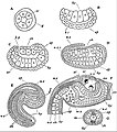
English: Stages in the Embryology of a Simple Ascidian. See legend below.
Date published 1911
Source “Tunicata,” Encyclopædia Britannica (11th ed.), v. 27, 1911, p. 384, fig. 13.
Author After Kowalevsky.
Permission
(Reusing this file)
Public domain This image comes from the 13th edition of the Encyclopædia Britannica or earlier. The copyrights for that book have expired in the United States because the book was first published in the US with the publication occurring before January 1, 1929. As such, this image is in the public domain in the United States.
English: Legend:

A to F, Longitudinal vertical sections of embryos, all placed with the dorsal surface uppermost and the anterior end at the right.

A, Early blastula stage, during segmentation.

B, Early gastrula stage.

C, Stage after gastrula, showing commencement of notochord.

D, Later stage, showing formation of notochord and of neural canal.

E, Embryo showing body and tail and completely formed neural canal.

F, Larva just hatched; end of tail cut off.

G, Transverse section of tail of larva.

adp, Adhering papillae of larva.
at, Epiblastic (atrial) involution.
au, Auditory organ of larva.
ar, Archenteron.
bc, Blastocoele.
bp, Blastopore.
ch, Notochord.
ep, Epiblast.
hy, Hypoblast.
nc, Neural canal.
nec, Neurenteric canal.
oc, Ocular organ of larva.
g, Gelatinous investment of embryo.
m, Muscle cells of tail.
mes, Mesenteron.
mc, Mesoderm cells.
nv, Cerebral vesicle at anterior end of neural canal.

Captions

Add a one-line explanation of what this file represents

Items portrayed in this file

depicts

image/jpeg

File history

Click on a date/time to view the file as it appeared at that time.

Date/TimeThumbnailDimensionsUserComment
current19:42, 17 July 2019Thumbnail for version as of 19:42, 17 July 2019805 × 913 (216 KB)Bob Burkhardt{{Information |description ={{en|1=Stages in the Embryology of a Simple Ascidian. See legend below.}} |date =published 1911 |source =“Tunicata,” ''Encyclopædia Britannica'' (11th ed.), v. 27, 1911, p. 384, fig. 13. |author =After Kowalevsky. |permission ={{PD-Britannica}} }} {{en|Legend:}} A to F, Longitudinal vertical sections of embryos, all placed with the dorsal surface uppermost and the anterior end at the right. A, Early blastula stage, during segmentation....