Jhering) that they are truly processes of the head. It appears to be impossible to doubt that the lobes in question are the fore-portion of the foot, when their development is examined (see fig. 35), further, when the fact is considered that they are innervated by the pedal ganglion.
 |
| Fig. 5.—View of the postero-ventral surface of a male Pearly Nautilus, the mantle-skirt (c) being completely reflected so as to show the inner wall of the sub-pallial chamber, and the four ctenidia and the foot cut short (drawn from nature by A. G. Bourne). pe, Penis, being the enlarged termination of the right spermatic duct; l.sp, aperture of the rudimentary left spermatic duct (pyriform sac of Owen). Other letters as in fig. 4. |
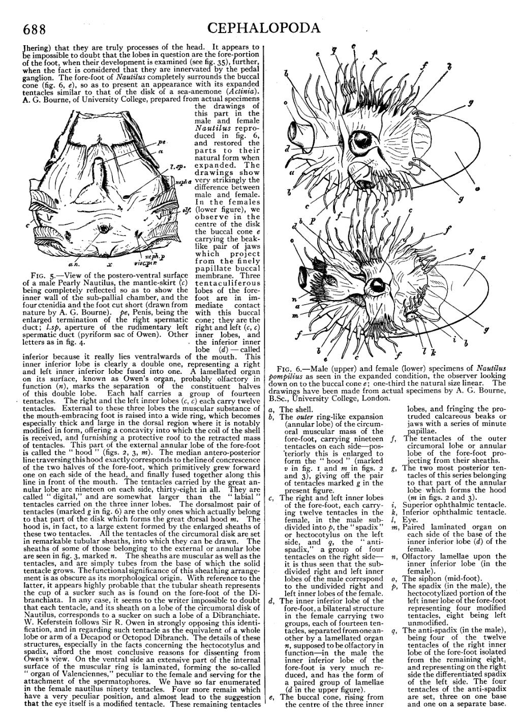
The fore-foot of Nautilus completely surrounds the buccal cone (fig. 6, e), so as to present an appearance with its expanded tentacles similar to that of the disk of a sea-anemone (Actinia). A. G. Bourne, of University College, prepared from actual specimens the drawings of this part in the male and female Nautilus reproduced in fig. 6, and restored the parts to their natural form when expanded. The drawings show very strikingly the difference between male and female. In the females (lower figure), we observe in the centre of the disk the buccal cone e carrying the beak-like pair of jaws which project from the finely papillate buccal membrane. Three tentaculiferous lobes of the fore-foot are in immediate contact with this buccal cone; they are the right and left (c, c) inner lobes, and the inferior inner lobe (d)—called inferior because it really lies ventralwards of the mouth. This inner inferior lobe is clearly a double one, representing a right and left inner inferior lobe fused into one. A lamellated organ on its surface, known as Owen’s organ, probably olfactory in function (n), marks the separation of the constituent halves of this double lobe. Each half carries a group of fourteen tentacles. The right and the left inner lobes (c, c) each carry twelve tentacles. External to these three lobes the muscular substance of the mouth-embracing foot is raised into a wide ring, which becomes especially thick and large in the dorsal region where it is notably modified in form, offering a concavity into which the coil of the shell is received, and furnishing a protective roof to the retracted mass of tentacles. This part of the external annular lobe of the fore-foot is called the “hood” (figs. 2, 3, m). The median antero-posterior line traversing this hood exactly corresponds to the line of concrescence of the two halves of the fore-foot, which primitively grew forward one on each side of the head, and finally fused together along this line in front of the mouth. The tentacles carried by the great annular lobe are nineteen on each side, thirty-eight in all. They are called “digital,” and are somewhat larger than the “labial” tentacles carried on the three inner lobes. The dorsalmost pair of tentacles (marked g in fig. 6) are the only ones which actually belong to that part of the disk which forms the great dorsal hood m. The hood is, in fact, to a large extent formed by the enlarged sheaths of these two tentacles. All the tentacles of the circumoral disk are set in remarkable tubular sheaths, into which they can be drawn. The sheaths of some of those belonging to the external or annular lobe are seen in fig. 3, marked n. The sheaths are muscular as well as the tentacles, and are simply tubes from the base of which the solid tentacle grows. The functional significance of this sheathing arrangement is as obscure as its morphological origin. With reference to the latter, it appears highly probable that the tubular sheath represents the cup of a sucker such as is found on the fore-foot of the Dibranchiata. In any case, it seems to the writer impossible to doubt that each tentacle, and its sheath on a lobe of the circumoral disk of Nautilus, corresponds to a sucker on such a lobe of a Dibranchiate. W. Keferstein follows Sir R. Owen in strongly opposing this identification, and in regarding such tentacle as the equivalent of a whole lobe or arm of a Decapod or Octopod Dibranch. The details of these structures, especially in the facts concerning the hectocotylus and spadix, afford the most conclusive reasons for dissenting from Owen’s view. On the ventral side an extensive part of the internal surface of the muscular ring is laminated, forming the so-called “organ of Valenciennes,” peculiar to the female and serving for the attachment of the spermatophores. We have so far enumerated in the female nautilus ninety tentacles. Four more remain which have a very peculiar position, and almost lead to the suggestion that the eye itself is a modified tentacle. These remaining tentacles are placed one above (before) and one below (behind) each eye, and bring up the total to ninety-four (fig. 3 v, v).
_and_female_(lower)_specimens_of_Nautilus_pompilius.jpg) | |
| Fig. 6.—Male (upper) and female (lower) specimens of Nautilus pompilius as seen in the expanded condition, the observer looking down on to the buccal cone e; one-third the natural size linear. The drawings have been made from actual specimens by A. G. Bourne, B.Sc., University College, London. | |
a, The shell.
b, The outer ring-like expansion (annular lobe) of the circumoral muscular mass of the fore-foot, carrying nineteen tentacles on each side—posteriorly this is enlarged to form the “hood” (marked v in fig. 1 and m in figs. 2 and 3). giving off the pair of tentacles marked g in the present figure.
c, The right and left inner lobes of the fore-foot, each carrying twelve tentacles in the female, in the male subdivided into p, the “spadix” or hectocotylus on the left side, and q, the “anti-spadix,” a group of four tentacles on the right side—it is thus seen that the subdivided right and left inner lobes of the male correspond to the undivided right and left inner lobes of the female.
d, The inner inferior lobe of the fore-foot, a bilateral structure in the female carrying two groups, each of fourteen tentacles, separated from one another by a lamellated organ n, supposed to be olfactory in function—in the male the inner inferior lobe of the fore-foot is very much reduced, and has the form of a paired group of lamellae (d in the upper figure).
e, The buccal cone, rising from the centre of the three inner lobes, and fringing the protruded calcareous beaks or jaws with a series of minute papillae. |
f,The tentacles of the outer circumoral lobe or annular lobe of the fore-foot projecting from their sheaths.
g, The two most posterior tentacles of this series belonging to that part of the annular lobe which forms the hood (m in figs. 2 and 3).
i,Superior ophthalmic tentacle.
k,Inferior ophthalmic tentacle.
l,Eye.
m, Paired laminated organ on each side of the base of the inner inferior lobe (d) of the female.
n, Olfactory lamellae upon the inner inferior lobe (in the female).
o, The siphon (mid-foot).
p, The spadix (in the male), the hectocotylized portion of the left inner lobe of the fore-foot representing four modified tentacles, eight being left unmodified.
q, The anti-spadix (in the male), being four of the twelve tentacles of the right inner lobe of the fore-foot isolated from the remaining eight, and representing on the right side the differentiated spadix of the left side. The four tentacles of the anti-spadix are set, three on one base and one on a separate base. |