the special student of the Crustacea, and cannot be fully dealt with here.
Segmentation is usually of the superficial or centrolecithal type. The hypoblast is formed either by a definite invagination or by the immigration of isolated cells, known as vitellophags, which wander through the yolk and later become associated into a definite mesenteron, or by some combination of these two methods. The blastopore generally occupies a position corresponding to the posterior end of the body. The mesoblast of the cephalic (naupliar) region probably arises in connexion with the lips of the blastopore and consists of loosely-connected cells or mesenchyme. In the region of the trunk, in many cases, paired mesoblastic bands are formed, growing in length by the division of teloblastic cells at the posterior end, and becoming segmented into somites. The existence of true coelom-sacs is somewhat doubtful. The rudiments of the first three pairs of appendages commonly appear simultaneously, and, even in forms with embryonic development, they show differences in their mode of appearance from the succeeding somites. Further, a definite cuticular membrane is frequently formed and shed at this stage, which corresponds to the nauplius-stage of larval development.
 |
| Fig. 12.—Nauplius of a Prawn (Penaeus). (Fritz Müller). |
The larval metamorphoses of the Crustacea have attracted much attention, and have been the subject of much discussion in view of their bearing on the phylogenetic history of the group. In those Crustacea in which the series of larval stages is most complete, the starting-point is the form already mentioned under the name of nauplius. The typical nauplius (fig. 12) has an oval unsegmented body and three pairs of limbs corresponding to the antennules, antennae and mandibles of the adult. The antennules are uniramous, the others biramous, and all three pairs are used in swimming. The antennae have a spiniform or hooked masticatory process at the base, and share with the mandibles, which have a similar process, the function of seizing and masticating the food. The mouth is overhung by a large labrum or upper lip, and the integument of the dorsal surface of the body forms a more or less definite dorsal shield. The paired eyes are, as yet, wanting, but the unpaired eye is large and conspicuous. A pair of frontal papillae or filaments, probably sensory, are commonly present.
A nauplius larva differing only in details from the typical form just described is found in the majority of the Phyllopoda, Copepoda and Cirripedia, and in a more modified form, in some Ostracoda. Among the Malacostraca the nauplius is less commonly found, but it occurs in the Euphausiidae among the Schizopoda and in a few of the more primitive Decapoda (Penaeidea) (fig. 12). In most of the Crustacea which hatch at a later stage there is, as already mentioned, more or less clear evidence of an embryonic nauplius stage. It seems certain, therefore, that the possession of a nauplius larva must be regarded as a very primitive character of the Crustacean stock.
As development proceeds, the body of the nauplius elongates, and indications of segmentation begin to appear in its posterior part. At successive moults the somites increase in number, new somites being added behind those already differentiated, from a formative zone in front of the telsonic region. Very commonly the posterior end of the body becomes forked, two processes growing out at the sides of the anus and often persisting in the adult as the “caudal furca.” The appendages posterior to the mandibles appear as buds on the ventral surface of the somites, and in the most primitive cases they become differentiated, like the somites which bear them, in regular order from before backwards. The limb-buds early become bilobed and grow out into typical biramous appendages which gradually assume the characters found in the adult. With the elongation of the body, the dorsal shield begins to project posteriorly as a shell-fold, which may increase in size to envelop more or less of the body or may disappear altogether. The rudiments of the paired eyes appear under the integument at the sides of the head, but only become pedunculated at a comparatively late stage.
The course of development here outlined, in which the nauplius gradually passes into the adult form by the successive addition of somites and appendages in regular order, agrees so well with the process observed in the development of the typical Annelida that we must regard it as being the most primitive method. It is most closely followed by the Phyllopods such as Apus or Branchipus, and by some Copepoda.
 | |
| Fig. 14.—Zoea of Common Shore-Crab in its second stage. (Spence Bate.) | |
| r, Rostral spine. | t, Buds of thoracic feet. |
| s, Dorsal spine. | m, Maxillipeds. |
| a, Abdomen. | |
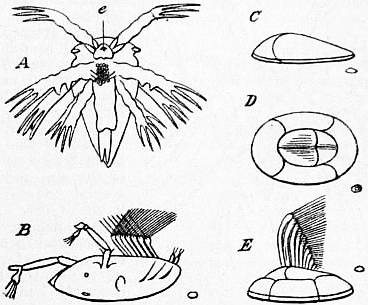
In most Crustacea, however, this primitive scheme is more or less modified. The earlier stages may be suppressed or passed through within the egg (or within the maternal brood-chamber), so that the larva, on hatching, has reached a stage more advanced than the nauplius. Further, the gradual appearance and differentiation of the successive somites and appendages may be accelerated, so that comparatively great advances take place at a single moult. In the Cirripedia, for example, the latest nauplius stage (fig. 13, A) gives rise directly to the so-called Cypris-larva (fig. 13, B), differing widely from the nauplius in form, and possessing all the appendages of the adult. Another very common modification of the primitive method of development is found in the accelerated appearance of certain somites or appendages, disturbing the regular order of development. This modification is especially found in the Malacostraca. Even in those which have most fully retained the primitive order of development, as in the Penaeidea and Euphausiidae, the last pair of abdominal appendages make their appearance in advance of those immediately in front of them. The same process, carried further, leads to the very peculiar larva known as the Zoea, in the typical form of which, found in the Brachyura (fig. 14), the posterior five or six thoracic somites have their development greatly retarded, and are still represented by a short unsegmented region of the body at a time when the abdominal somites are fully formed and even carry appendages. The Zoea was formerly regarded as a recapitulation of an ancestral form, but there can be no doubt that its peculiarities are the result of secondary modification. It is most typically developed in the most specialized Decapoda, the Brachyura, while the more primitive groups of Malacostraca, the Euphausiidae, Penaeidea and Stomatopoda, retain the primitive order of appearance

