vertebrae vary from ten in some of the whales and the peba armadillo to twenty-four in the two- toed sloth, though thirteen or fourteen is the commonest number. In the anterior part of the thoracic region the spines point backward, while in the posterior thoracic and lumbar regions they have a forward direction. There is always one spine in the posterior thoracic region, which is vertical, and the vertebra which bears this is known as the anticlinal vertebra. The
az
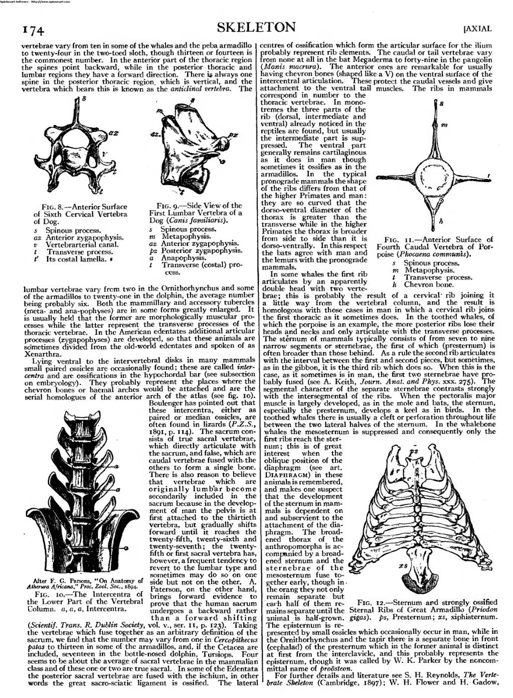
Fig. 8. — Anterior Surface of Sixth Cervical Vertebra of Dog.
s Spinous process. az Anterior zygapophysis. v Vertebrarterial canal. t Transverse process. V Its costal lamella. «
Fig. 9. — Side View of the First Lumbar Vertebra of a Dog {Canis familiaris). s Spinous process. m Metapophysis. az Anterior zygapophysis. pz Posterior zygapophysis. a Anapophysis. / Transverse (costal) pro- cess.
lumbar vertebrae vary frcm two in the Omithorhynchus and some of the armadillos to twenty-one in the dolphin, the average number being probably six. Both the mammillary and accessory tubercles (meta- and ana-pophyses) are in some forms greatly enlarged. It is usually held that the former are morphologically muscular pro- cesses while the latter represent the transverse processes of the thoracic vertebrae. In the American edentates additional articular processes (zygapophyses) are developed, so that these animals are sometimes divided from the old-world edentates and spoken of as Xenarthra.
Lying ventral to the intervertebral disks in many mammals small paired ossicles are occasionally found; these are called inter- centra and are ossifications in the hypochordal bar (see subsection on embryology). They probably represent the places where the chevron bones or haemal arches would be attached and are the serial homologues of the anterior arch of the atlas (see fig. 10).
Boulenger has pointed out that these intercentra, either as paired or median ossicles, are often found in lizards (P.Z.S., i8<}i,p. 114). The sacrum con- sists of true sacral vertebrae, which directly articulate with the sacrum, and false, which are caudal vertebrae fused with- the others to form a single bone. There is also reason to believe that vertebrae which are originally lumbar become secondarily included in the sacrum because in the develop- ment of man the pelvis is at first attached to the thirtieth vertebra, but gradually shifts forward until it reaches the twenty -fifth, twenty-sixth and twenty-seventh ; the twenty- fifth or first sacral vertebra has, however, a frequent tendency to revert to the lumbar type and sometimes may do so on one side but not on the other. A. Paterson, on the other hand, brings forward evidence to prove that the human sacrum undergoes a backward rather than a forward shifting (Scientif. Trans. R. Dublin Society, vol. v., ser. 11, p. 123). Taking the vertebrae which fuse together as an arbitrary definition of the sacrum, we find that the number may vary from one in Cercopithecus patas to thirteen in some of the armadillos, and, if the Cetacea are included, seventeen in the bottle-nosed dolphin, Tursiops. Four seems to be about the average of sacral vertebrae in the mammalian class and of these one or two are true sacral. In some of the Edentata the posterior sacral vertebrae are fused with the ischium, in other words the great sacro-sciatic ligament is ossified. The lateral
After F. G. Parsons, "On Anatomy of
Alherura Africana," Proc. Zool. Soc, 1894.
Fig. 10. — The Intercentra of the Lower Part of the Vertebral Column, a, a, a, Intercentra.
centres of ossification which form the articular surface for the ilium probably represent rib elements. The caudal or tail vertebrae vary from none at all in the bat Megaderma to forty-nine in the pangolin {Manis macrura). The anterior ones are remarkable for usually having chevron bones (shaped like a V) on the ventral surface of the intercentral articulation. These protect the caudal vessels and give attachment to the ventral tail muscles. The ribs in mammals correspond in number to the thoracic vertebrae. In mono- tremes the three parts of the rib (dorsal, intermediate and ventral) already noticed in the reptiles are found, but usually the intermediate part is sup- pressed. The ventral part generally remains cartilaginous as it does in man though sometimes it ossifies as in the armadillos. In the typical pronograde mammals the shape of the ribs differs from that of the higher Primates and man : they are so curved that the dorso-ventral diameter of the thorax is greater than the transverse while in the higher Primates the thorax is broader from side to side than it is dorso-ventrally. In this respect the bats agree with man and the lemurs with the pronograde mammals.
In some whales the first rib articulates by an apparently double head with two verte- brae; this is probably the result little way from the vertebral
Fig. ii. — Anterior Surface of
Fourth Caudal Vertebra of Por-
poise (Phocaena communis),
s Spinous process.
Metapophysis.
Transverse process.
Chevron bone.
m
t
h
of a cervical " column, and
rib joining the result
homologous with those cases in man in which a cervical rib joins the first thoracic as it sometimes does. In the toothed whales, of which the porpoise is an example, the more posterior ribs lose their heads and necks and only articulate with the transverse processes. The sternum of mammals typically consists of from seven to nine narrow segments or sternebrae, the first of which (presternum) is often broader than those behind. As a rule the second rib articulates with the interval between the first and second pieces, but sometimes, as in the gibbon, it is the third rib which does so. When this is the case, as it sometimes is in man, the first two sternebrae have pro- bably fused (see A. Keith, Journ. Anat. and Phys. xxx. 275). The segmental character of the separate sternebrae contrasts strongly with the intersegmental of the ribs. When the pectoralis major muscle is largely developed, as in the mole and bats, the sternum, especially the presternum, develops a keel as in birds. In the toothed whales there is usually a cleft or perforation throughout life between the two lateral halves of the sternum. In the whalebone whales the mesosternum is suppressed and consequently only the first ribs reach the ster- num; this is of great interest when the oblique position of the diaphragm (see art. Diaphragm) in these animals is remembered, and makes one suspect that the development of the sternum in mam- mals is dependent on and subservient to the attachment of the dia- phragm. The broad- ened thorax of the anthropomorpha is ac- companied by a broad- ened sternum and the sternebrae of the mesosternum fuse to- gether early, though in the orang they not only remain separate but each half of them re- mains separate until the animal is half -grown. The episternum is re- presented by small ossicles which occasionally occur in man, while in the Omithorhynchus and the tapir there is a separate bone in front (cephalad) of the presternum which in the former animal is distinct at first from the interclavicle, and this probably represents the episternum, though it was called by W. K. Parker by the noncom- mittal name of probsteon.
For further details and literature see S. H. Reynolds, The Verte- brate Skeleton (Cambridge, 1897); W. H. Flower and H. Gadow,
An image should appear at this position in the text. To use the entire page scan as a placeholder, edit this page and replace "{{missing image}}" with "{{raw image|EB1911 - Volume 25.djvu/190}}". Otherwise, if you are able to provide the image then please do so. For guidance, see Wikisource:Image guidelines and Help:Adding images. |
Fig. 12. — Sternum and strongly ossified Sternal Ribs of Great Armadillo (Priodon gigas). ps, Presternum; xs, xiphisternum.