Medulla Oblongata.
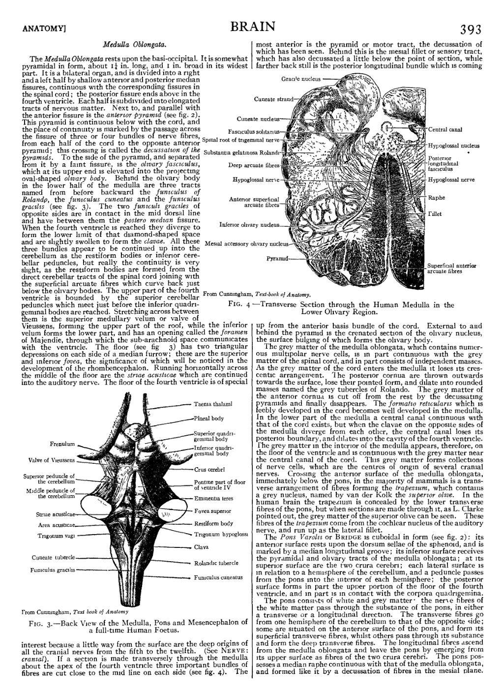
The Medulla Oblongata rests upon the basi-occipital. It is somewhat pyramidal in form, about 114 in. long, and 1 in. broad in its widest part. It is a bilateral organ, and is divided into a right and a left half by shallow anterior and posterior median fissures, continuous with the corresponding fissures in the spinal cord; the posterior fissure ends above in the fourth ventricle. Each half is subdivided into elongated tracts of nervous matter. Next to, and parallel with the anterior fissure is the anterior pyramid (see fig. 2). This pyramid is continuous below with the cord, and the place of continuity is marked by the passage across the fissure of three or four bundles of nerve fibres, from each half of the cord to the opposite anterior pyramid; this crossing is called the decussation of the pyramids. To the side of the pyramid, and separated from it by a faint fissure, is the olivary fasciculus, which at its upper end is elevated into the projecting oval-shaped olivary body. Behind the olivary body in the lower half of the medulla are three tracts named from before backward the funiculus of Rolando, the funiculus cuneatus and the funiculus gracilis (see fig. 3). The two funiculi graciles of opposite sides are in contact in the mid dorsal line and have between them the postero median fissure. When the fourth ventricle is reached they diverge to form the lower limit of that diamond-shaped space and are slightly swollen to form the clavae. All these three bundles appear to be continued up into the cerebellum as the restiform bodies or inferior cerebellar peduncles, but really the continuity is very slight, as the restiform bodies are formed from the direct cerebellar tracts of the spinal cord joining with the superficial arcuate fibres which curve back just below the olivary bodies. The upper part of the fourth ventricle is bounded by the superior cerebellar peduncles which meet just before the inferior quadrigeminal bodies are reached. Stretching across between them is the superior medullary velum or valve of Vieussens, forming the upper part of the roof, while the inferior velum forms the lower part, and has an opening called the foramen of Majendie, through which the sub-arachnoid space communicates with the ventricle. The floor (see fig. 3) has two triangular depressions on each side of a median furrow; these are the superior and inferior fovea, the significance of which will be noticed in the development of the rhombencephalon. Running horizontally across the middle of the floor are the striae acusticae which are continued into the auditory nerve. The floor of the fourth ventricle is of special interest because a little way from the surface are the deep origins of all the cranial nerves from the fifth to the twelfth. (See Nerve, cranial). If a section is made transversely through the medulla about the apex of the fourth ventricle three important bundles of fibres are cut close to the mid line on each side (see fig. 4). The most anterior is the pyramid or motor tract, the decussation of which has been seen. Behind this is the mesial fillet or sensory tract, which has also decussated a little below the point of section, while farther back still is the posterior longitudinal bundle which is coming up from the anterior basis bundle of the cord. External to and behind the pyramid is the crenated section of the olivary nucleus, the surface bulging of which forms the olivary body.

|
From Cunningham, Text-book of Anatomy. |
| Fig. 3.—Back View of the Medulla, Pons and Mesencephalon of a full-time Human Foetus. |
The grey matter of the medulla oblongata, which contains numerous multipolar nerve cells, is in part continuous with the grey matter of the spinal cord, and in part consists of independent masses. As the grey matter of the cord enters the medulla it loses its crescentic arrangement. The posterior cornua are thrown outwards towards the surface, lose their pointed form, and dilate into rounded masses named the grey tubercles of Rolando. The grey matter of the anterior cornua is cut off from the rest by the decussating pyramids and finally disappears. The formatio reticularis which is feebly developed in the cord becomes well developed in the medulla. In the lower part of the medulla a central canal continuous with that of the cord exists, but when the clavae on the opposite sides of the medulla diverge from each other, the central canal loses its posterior boundary, and dilates into the cavity of the fourth ventricle. The grey matter in the interior of the medulla appears, therefore, on the floor of the ventricle and is continuous with the grey matter near the central canal of the cord. This grey matter forms collections of nerve cells, which are the centres of origin of several cranial nerves. Crossing the anterior surface of the medulla oblongata, immediately below the pons, in the majority of mammals is a transverse arrangement of fibres forming the trapezium, which contains a grey nucleus, named by van der Kolk the superior olive. In the human brain the trapezium is concealed by the lower transverse fibres of the pons, but when sections are made through it, as L. Clarke pointed out, the grey matter of the superior olive can be seen. These fibres of the trapezium come from the cochlear nucleus of the auditory nerve, and run up as the lateral fillet.
From Cunningham, Text-book of Anatomy. |
| Fig. 4.—Transverse Section through the Human Medulla in the Lower Olivary Region. |
The Pons Varolii or Bridge is cuboidal in form (see fig. 2): its anterior surface rests upon the dorsum sellae of the sphenoid, and is marked by a median longitudinal groove; its inferior surface receives the pyramidal and olivary tracts of the medulla oblongata; at its superior surface are the two crura cerebri; each lateral surface is in relation to a hemisphere of the cerebellum, and a peduncle passes from the pons into the interior of each hemisphere; the posterior surface forms in part the upper portion of the floor of the fourth ventricle, and in part is in contact with the corpora quadrigemina.
The pons consists of white and grey matter: the nerve fibres of the white matter pass through the substance of the pons, in either a transverse or a longitudinal direction. The transverse fibres go from one hemisphere of the cerebellum to that of the opposite side; some are situated on the anterior surface of the pons, and form its superficial transverse fibres, whilst others pass through its substance and form the deep transverse fibres. The longitudinal fibres ascend from the medulla oblongata and leave the pons by emerging from its upper surface as fibres of the two crura cerebri. The pons possesses a median raphe continuous with that of the medulla oblongata, and formed like it by a decussation of fibres in the mesial plane.