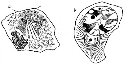
division into two of each of the chromatin granules. In the spermatogenic cells of Ascaris, A. Brauer has shown that the chromatin granules divide while still scattered over the nuclear reticulum and before either the formation of a spireme thread or the division of the centrosome. In many other cases the reverse of this condition occurs, the centrosome dividing long before there is any indication of division in the nucleus (e.g. salamander spermatogenic cells, Meves, &c.). We must therefore, with Boveri and Brauer, regard the division of the chromatin in mitosis as a distinct reproductive act on the part of the chromatin granules, the chromosomes being merely aggregates (temporary or permanent, vide infra) of these self-propagating units.
For convenience of description it is usual to recognize four periods in mitosis: (i.) Prophase, (ii.) Metaphase, (iii.) Anaphase, and (iv.) Telophase (Strasburger, 1884). The prophase covers all changes up to the completion of the mitotic figure. The metaphase is the parting of the sister chromosomes in the equatorial plate; their passage to opposite poles of the spindle constitutes the anaphase; and their reconstruction to form the resting daughter nuclei, the telophase.

Fig. 7.—Centrosomes.
From Prof. E. B. Wilson’s The Cell in Development and Inheritance, by permission of the author and of The Macmillan Co., New York.
a, Leucocyte from a Salamander, showing permanent aster and centrosome.
From A. Gurwitsch, Morphologie u. Biologie der Zelle, by permission of Gustav Fischer.
b, Sperm-mother cell of Salamandra maculata, showing Hermann’s “central spindle.”
The Achromatic Figure.—The mode of origin of the achromatic figure varies greatly. In some cases a distinct and continuous spindle, the “central spindle” of F. Hermann, is visible from the very first separation of the daughter centrosomes (e.g. salamander spermatogenic cell)[1] (fig. 7, b). In other cases the rays only invade the nuclear area and become continuous in the equatorial plane after the centrosomes have assumed their definitive positions at the two poles of the nucleus, and may even appear to indent the disappearing nuclear membrane as they invade the nuclear area.[2] In the salamander testis cell (fig. 7, b), and in many other cases, the whole of the achromatic figure is obviously of cytoplasmic origin. In many cases, however, it equally obviously arises within the nucleus,[3] while in yet other cases[4] the spindle fibres are of mixed origin. The question, therefore, of the cytoplasmic or nuclear origin of the achromatic figure, at one time regarded as of considerable importance, is wholly immaterial. Various elaborate theories have been propounded to explain the mechanism of the mitotic figure. H. Fol (1873) regarded the centrosomes as centres of attractive forces, and compared the mitotic figure to the lines of force in the magnetic field, a comparison made by numerous subsequent workers. E. Klein’s hypotheses of two opposing systems of contractile fibrillae, elaborated by van Beneden (1883, 1887) and accepted by Boveri (1888), was still further extended by R. Heidenhain in relation to the leucocytes of the salamander, in which there is a permanent centrosome and astral rays to which the contractile movements of the cell appear to be due[5] (fig. 7, a). Hermann on the other hand confined the contractility to the astral and mantle fibres; while L. Druner regarded the spindle as exerting a pushing force, for not only do the interzonal spindle fibres elongate during the anaphase, but they were often at this period contorted, while on the other hand astral rays may be entirely absent (e.g. Infusoria), and in some cases the spindle pole may be caused to project at the surface of the cell. The futility of these attempted mechanical explanations of mitosis is sufficiently clearly shown, not only by the contradictory nature of the explanations themselves, but by the fact that, in amitosis, nuclear and cytoplasmic division occur without any fibrillar mechanism whatever.
Centrosome.[6]—This minute body was first detected at the spindle poles by Flemming in 1875, and independently by P. J. van Beneden in 1876. The important part played by the centrosome in fertilization,[7] first described by van Beneden and Theodor Boveri in their papers of 1887–1888, together with the behaviour of this structure in mitosis, led these authors to regard the centrosome not only as the dynamic centre of the cell but as a permanent cell-organ, which, like the nucleus, passed by division from one cell-generation to the next. This conclusion appeared to receive considerable support from the recognition of the centrosome in various kinds of resting cells,[8] and especially from the relation this structure frequently shows to the locomotor apparatus of the cell (e.g. its position in the centre of the radiating fibrillae in the contractile lymph and pigment cells, and its relation to the vibratile flagellum in spermatozoa and some protozoa, e.g. Trypanosoma).[9] In almost all cases the centrosome of the resting cell, when this can be detected, lies in the cytoplasm, and is often already divided in preparation for the next mitotic division (e.g. spermatogenic cells of the salamander; Meves). In some cases, however, it resides in, or arises from, the nucleus (Brauer; spermatogenesis of Ascaris, var. univalens). This indifferent nuclear or cytoplasmic position for the centrosome is paralleled by the attraction sphere or homologue of the centrosome in many Protozoa. Thus in many forms, e.g. Euglena (Keuten), it lies within the nucleus, while in other forms, e.g. Noctiluca (Ishikawa, 1894, 1898; Calkins, 1898) and Paramoeba (F. Schaudinn, 1896), it lies in the cytoplasm, while in Tetramitus it coexists with a “distributed” nucleus. In the Heliozoa conditions are exceptionally interesting; not only is the centrosome—here resembling in appearance that of the higher forms—permanently visible and extranuclear, lying at the centre of the radiations characteristic of these forms, but there is the strongest possible evidence for its formation de novo. For Schaudinn has shown in Acanthocystis that, in the formation of the swarm spores, the nucleus divides amitotically, the centrosome remaining visible and unchanged at the centre of the radiating processes. Yet a centrosome appears later in the nucleus of the swarm spores and migrates into the cytoplasm. The experiments of T. H. Morgan and E. B. Wilson, in which numerous centrosomes and asters (“cytasters”) are caused to appear in unfertilized sea-urchin eggs by a brief immersion in a 13% solution of magnesium
- ↑ The discovery by Hermann of the central spindle first clearly showed that two kinds of fibres must be recognized in the mitotic figure. Those of the central spindle correspond to the continuous spindle fibres of Flemming (1891) and Strasburger (1884), and the mantle fibres, i.e. half-spindle or Polstrahlen, of van Beneden (1887) and Boveri (1889–1890).
- ↑ Planter, Watasé, Griffen and others.
- ↑ e.g. Euglypha (Schewiakoff, 1888), Infusoria (R. Hertwig, 1898). So also Korschelt for Ophryotrocha, and many other cases.
- ↑ e.g. Bauer, spermatogenic cells of Ascaris univalens.
- ↑ Cf. also Watasé, Solger and Zimmermann.
- ↑ This term is due to Boveri (Zellenstudien, ii., 1888, p. 68; Jen. Zeit. xxii.), but it was intended by him to include the region of modified cytoplasm or “centrosphere” often enclosing the centrosome proper, i.e. “centriole” of Boveri.
- ↑ For outline of fertilization see article Reproduction.
- ↑ e.g. lymph and various epithelial and connective tissue cells of salamander larva (Flemming, 1891; Heidenhain, 1892); pigment cells of fishes (Solger, 1891); red blood corpuscles (Heidenhain, Eisen, 1897); and numerous other cases.
- ↑ For an interesting development of this subject see Watasé (1894). This author not only identifies the centrosome with the structures seen in lymph cells, &c., but compares it to the basal granules of ciliated cells and to the varicose swellings on the sarcostyles of striped muscle cells!
