rhombencephalon (see Brain), and the sensory are the axons and dendrites of cells situated in ganglia which have budded off from the brain. The evidence of comparative anatomy, however, shows that the cranial nerves cannot be directly homologized with the spinal, nor can the fact of there being twelve of them justify us in assuming that the head contains the rudiments of twelve fused or unsegmented somites. To this we will return later. The case of the optic nerve is different to that of any of the others. A. Robinson (Journ. Anat. and Phys., vol. 30, p. 319) has shown that most of its fibres are the axons of ganglion cells in the retina, and, as the retina is part of the optic vesicle and an outgrowth from the brain, the so-called optic nerve is only comparable to a tract of fibres within the brain.
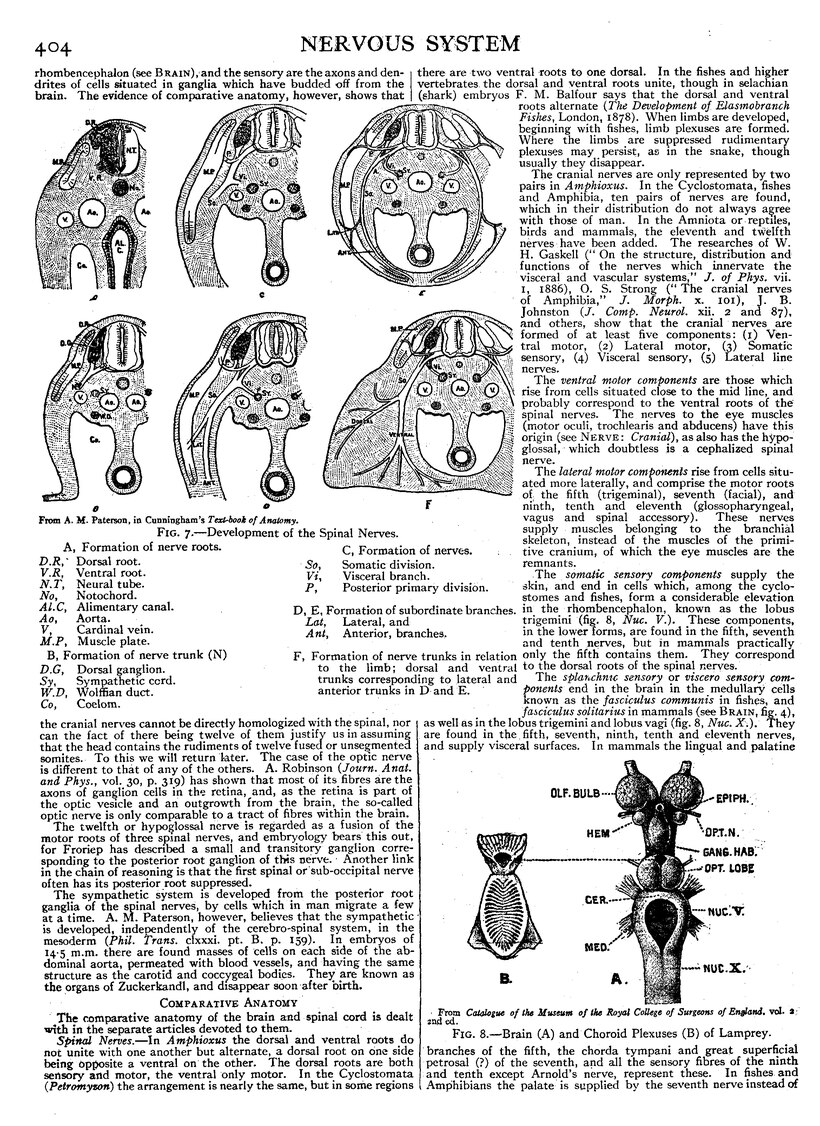
| From A. M. Paterson, in Cunningham’s Text-Book of Anatomy. | |||
| Fig. 7.—Development of the Spinal Nerves. | |||
| A, Formation of nerve roots. | C, Formation of nerves. | ||
| D.R, | Dorsal root. | So, | Somatic division. |
| V.R, | Ventral root. | Vi, | Visceral branch. |
| N.T, | Neural tube. | P, | Posterior primary division. |
| No, | Notochord. | ||
| Al.C, | Alimentary canal. | D, E, Formation of subordinate branches. | |
| Ao, | Aorta. | Lat, | Lateral, and |
| V, | Cardinal vein. | Ant, | Anterior, branches. |
| M.P, | Muscle plate. | ||
| B, Formation of nerve trunk (N). | F, Formation of nerve trunks in relation to the limb; dorsal and ventral trunks corresponding to lateral and anterior trunks in D and E.
| ||
| D.G, | Dorsal ganglion. | ||
| Sy, | Sympathetic cord. | ||
| W.D, | Wolffian duct. | ||
| Co, | Coelom. | ||
The twelfth or hypoglossal nerve is regarded as a fusion of the motor roots of three spinal nerves, and embryology bears this out, for Froriep has described a small and transitory ganglion corresponding to the posterior root ganglion of this nerve. Another link in the chain of reasoning is that the first spinal or sub-occipital nerve often has its posterior root suppressed.
The sympathetic system is developed from the posterior root ganglia of the spinal nerves, by cells which in man migrate a few at a time. A. M. Paterson, however, believes that the sympathetic is developed, independently of the cerebro-spinal system, in the mesoderm (Phil. Trans. clxxxi. pt. B. p. 159). In embryos of 14·5 m.m. there are found masses of cells on each side of the abdominal aorta, permeated with blood vessels, and having the same structure as the carotid and coccygeal bodies. They are known as the organs of Zuckerkandl, and disappear soon after birth.
Comparative Anatomy
The comparative anatomy of the brain and spinal cord is dealt with in the separate articles devoted to them.
Spinal Nerves.—In Amphioxus the dorsal and ventral roots do not unite with one another but alternate, a dorsal root on one side being opposite a ventral on the other. The dorsal roots are both sensory and motor, the ventral only motor. In the Cyclostomata (Petromyzon) the arrangement is nearly the same, but in some regions there are two ventral roots to one dorsal. In the fishes and higher vertebrates. the dorsal and ventral roots unite, though in selachian (shark) embryos F. M. Balfour says that the dorsal and ventral roots alternate (The Development of Elasmobranch Fishes, London, 1878). When limbs are developed, beginning with fishes, limb plexuses are formed. Where the limbs are suppressed rudimentary plexuses may persist as in the snake, though usually they disappear.
The cranial nerves are only represented by two pairs in Amphioxus. In the Cyclostomata, fishes and Amphibia, ten pairs of nerves are found, which in their distribution do not always agree with those of man. In the Amniota or reptiles, birds and mammals, the eleventh and twelfth nerves have been added. The researches of W. H. Gaskell (“On the structure, distribution and functions of the nerves which innervate the visceral and vascular systems,” J. of Phys. vii. 1, 1886), O. S. Strong (“The cranial nerves of Amphibia,” J. Morph. x. 101), J. B. Johnston (J. Comp. Neurol. xii. 2 and 87), and others, show that the cranial nerves are formed of at least five components: (1) Ventral motor, (2) Lateral motor, (3) Somatic sensory, (4) Visceral sensory, (5) Lateral line nerves.
The ventral motor components are those which rise from cells situated close to the mid line, and probably correspond to the ventral roots of the spinal nerves. The nerves to the eye muscles (motor oculi, trochlearis and abducens) have this origin (see Nerve: Cranial), as also has the hypoglossal, which doubtless is a cephalized spinal nerve.
The lateral motor components rise from cells situated more laterally, and comprise the motor roots of the fifth (trigeminal), seventh (facial), and ninth, tenth and eleventh (glossopharyngeal, vagus and spinal accessory). These nerves supply muscles belonging to the branchial skeleton, instead of the muscles of the primitive cranium, of which the eye muscles are the remnants.
The somatic sensory components supply the skin, and end in cells which, among the cyclostomes and fishes, form a considerable elevation in the rhombencephalon, known as the lobus trigemini (fig. 8, Nuc. V.). These components, in the lower forms, are found in the fifth, seventh and tenth nerves, but in mammals practically only the fifth contains them. They correspond to the dorsal roots of the spinal nerves.
| From Catalogue of the Museum of the Royal College of Surgeons of England, vol. 2, 2nd ed. |
| Fig. 8.—Brain (A) and Choroid Plexuses (B) of Lamprey. |
The splanchnic sensory or viscero sensory components end in the brain in the medullary cells known as the fasciculus communis in fishes, and fasciculus solitarius in mammals (see Brain, fig. 4), as well as in the lobus trigemini and lobus vagi (fig. 8, Nuc. X). They are found in the fifth, seventh, ninth, tenth and eleventh nerves, and supply visceral surfaces. In mammals the lingual and palatine branches of the fifth, the chorda tympani and great superficial petrosal (?) of the seventh, and all the sensory fibres of the ninth and tenth except Arnold's nerve, represent these. In fishes. and Amphibians the palate' is supplied by the seventh nerve instead of
