another, as in Charybdaea (fig. 4), where there are four gastric pouches
communicating with the central stomach by four so-called gastric
ostia (fig. 4). A similar condition is seen in Pelagia, where the
number of gastric pouches is
increased to sixteen. In forms
such as Lucernaria and Charybdaea,
in which the umbrella is
of deep form and the stomach-cavity
consequently of great
extent in the vertical direction,
the concrescence-areas or septal
nodes are drawn out into
vertical partitions or taeniolae
(fig. 4, L.o.c.), resembling in
their anatomical relations the
mesenteries of the Anthopolyp.
The phacellae are carried on
the edges of the taeniolae
(fig. 4, Gh). Finally in the
majority of Scyphomedusae
the primitively simple
concrescence-areas become
increased in number and in
extent, so that radial canals,
ring-canals, &c., can be distinguished
in addition to
stomach-pouches. Thus in Aurelia (figs.
2a and 2b), to take a familiar
example, the digestive tract
begins with the mouth, of
which the four corners are
prolonged into the four long
oral arms, perradial in position.
The mouth leads into the
spacious stomach containing
the four conspicuous
horse-shoe-shaped gonads (ov) marking
four stomach-pouches,
which, however, are interradial
in position. From the
stomach or its pouches arise
sixteen radial canals, four
perradial, four interradial and
eight adradial (fig. 2b). The
perradial and interradial canals
consist of a main stem giving
off branches, and both stem and branches reach to the marginal
ring-canal, the main stem ending in one of the eight tentaculocysts,
which are lodged in the notches between the lobes of the umbrellar
margin. The adradial canals are unbranched and run to the middle
point of one of the marginal lobes. The system of canals shows
great variation even in the same species.

| |
|
Fig. 2b.—Half of the lower surface of Aurelia aurita. The transparent tissues allow the enteric cavities and canals to be seen through them. (From Gegenbaur.) | |
| a, | Marginal lappets hiding tentaculocysts. |
| b, | Oral arms. |
| t, | tentacles. |
| v, | Axial or gastric portion of the enteric cavity. |
| gv, | Radiating and anastomosing canals of the enteric system. |
| ov, | Ovaries. The gastral filaments near to these are not drawn. |
The muscular system of the Scyphomedusae is developed on the subumbral surface as a system of circularly disposed fibres which by their contraction make the umbrella more concave and diminish its cavity. The circular muscles usually form two chief portions, a peripheral wreath-muscle (Kranzmuskel), subdivided into four, eight or sixteen areas, and an oral ring-muscle round the mouth. Endodermal muscles are found in the phacellae, and in such forms as Lucernaria, longitudinal (vertical) muscular tracts or bands are found in the taeniolae, which, according to some authorities, are of endodermal origin, but which, according to recent observations, are formed in the walls of the infundibular cavities, and are therefore of ectodermal origin.

|
|
Fig. 3.—Scyphomedusae. a, Rhizostoma pulmo; b, Chrysaora hysoscella. |

| |
|
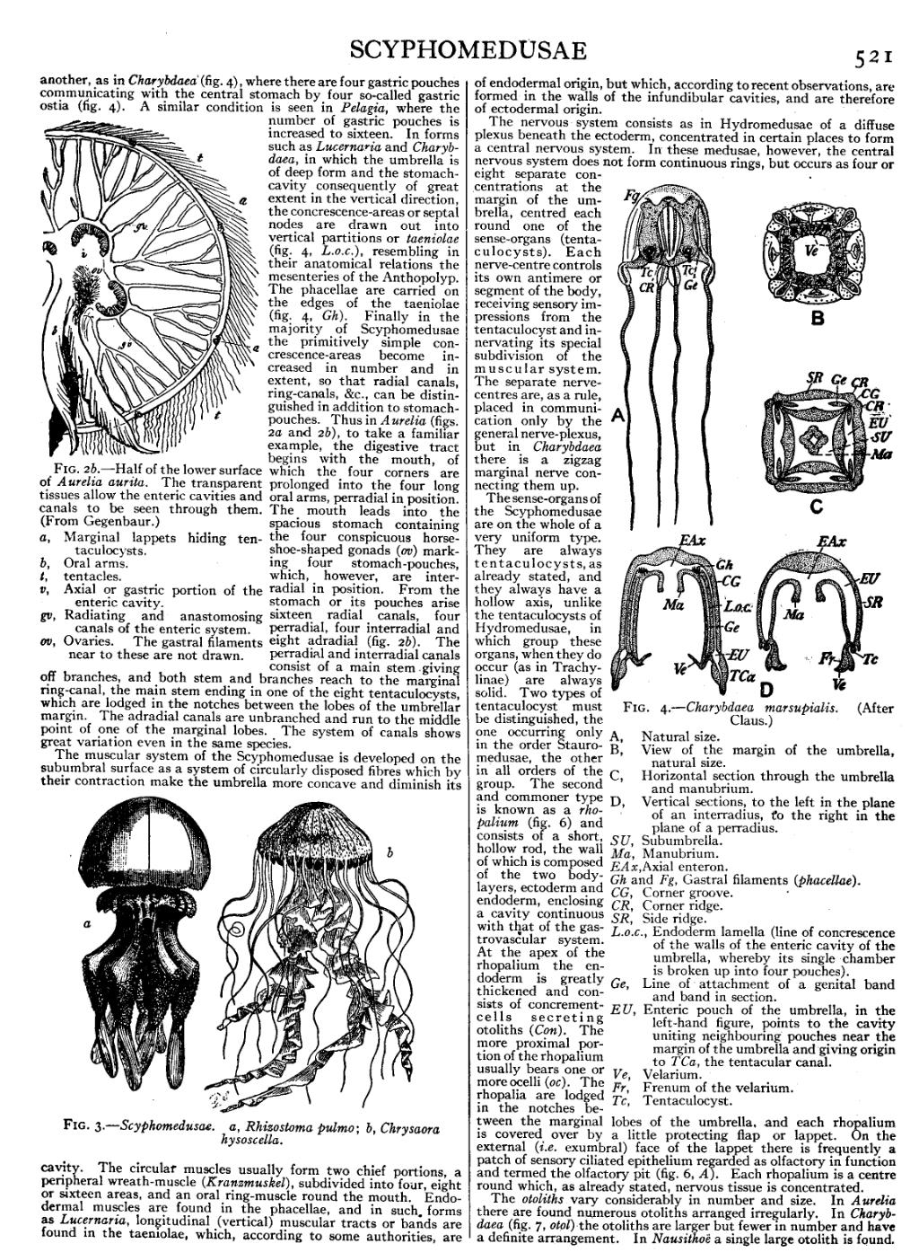
Fig. 4.—Charybdaea marsupialis. (After Claus.) | |
| A, | Natural size. |
| B, | View of the margin of the umbrella, natural size. |
| C, | Horizontal section through the umbrella and manubrium. |
| D, | Vertical sections, to the left in the plane of an interradius, to the right in the plane of a perradius. |
| SU, | Subumbrella. |
| Ma, | Manubrium. |
| EAx, | Axial enteron. |
| Gh and Fg, Gastral filaments (phacellae). | |
| CG, | Corner groove. |
| CR, | Corner ridge. |
| SR, | Side ridge. |
| L.o.c., | Endoderm lamella (line of concrescence of the walls of the enteric cavity of the umbrella, whereby its single chamber is broken up into four pouches). |
| Ge, | Line of attachment of a genital band and band in section. |
| EU, | Enteric pouch of the umbrella, in the left-hand figure, points to the cavity uniting neighbouring pouches near the margin of the umbrella and giving origin to TCa, the tentacular canal. |
| Ve, | Velarium. |
| Fr, | Frenum of the velarium. |
| Tc, | Tentaculocyst. |
The nervous system consists as in Hydromedusae of a diffuse plexus beneath the ectoderm, concentrated in certain places to form a central nervous system. In these medusae, however, the central nervous system does not form continuous rings, but occurs as four or eight separate concentrations at the margin of the umbrella, centred each round one of the sense-organs (tentaculocysts). Each nerve-centre controls its own antimere or segment of the body, receiving sensory impressions from the tentaculocyst and innervating its special subdivision of the muscular system. The separate nerve-centres are, as a rule, placed in communication only by the general nerve-plexus, but in Charybdaea there is a zigzag marginal nerve connecting them up.
The sense-organs of the Scyphomedusae are on the whole of a very uniform type. They are always tentaculocysts, as already stated, and they always have hollow axis, unlike the tentaculocysts of Hydromedusae, in which group these organs, when they do occur (as in Trachylinae) are always solid. Two types of tentaculocyst must be distinguished, the one occurring only in the order Stauromedusae, the other in all orders of the group. The second and commoner type is known as a rhopalium (fig. 6) and consists of a short, hollow rod, the wall of which is composed of the two body-layers, ectoderm and endoderm, enclosing a cavity continuous with that of the gastrovascular system. At the apex of the rhopalium the endoderm is greatly thickened and consists of concrement-cells secreting otoliths (Con). The more proximal portion of the rhopalium usually bears one or more ocelli (oc). The rhopalia are lodged in the notches between the marginal lobes of the umbrella, and each rhopalium is covered over by a little protecting flap or lappet. On the external (i.e. exumbral) face of the lappet there is frequently a patch of sensory ciliated epithelium regarded as olfactory in function and termed the olfactory pit (fig. 6, A). Each rhopalium is a centre round which, as already stated, nervous tissue is concentrated.
The otoliths vary considerably in number and size. In Aurelia there are found numerous otoliths arranged irregularly. In Charybdaea (fig. 7, otol) the otoliths are larger but fewer in number and have a definite arrangement. In Nausithoë a single large otolith is found.