Popular Science Monthly/Volume 6/January 1875/Biology for Young Beginners I
| BIOLOGY FOR YOUNG BEGINNERS.[1] |
By SARAH HACKETT STEVENSON.
UNDER the low eaves at the back of the house was a long, deep wooden trough for catching the rain that fell on the roof. This old trough was to me a never-failing source of wonder and delight during my childhood. The inside of it was all lined with a beautiful, green, velvety mould, and, when there had been no rain for some time, the water itself would turn a greenish color. We used to catch our little downy yellow ducks and put them in the trough to see them swim, and sometimes they would break off and eat the green mould with their curious shovel-bills. What this queer, green stuff was, and how it came there, was a great mystery to us children. Charley declared it came down in the rain just as the angle-worms that he used for fish-bait. I had to wait a long time to find some one to explain to me all about these simple things. No doubt I might have learned about them here at home, if I had tried hard enough; but it so happened that I found a great professor in London, who was teaching his students just what I wanted to know, and he explained so well what I had seen in the old water-trough, and many other curious things, that I have thought my young friends might like to hear about them also. I am sure I should have been very glad if I could have found any one to explain them to me when I was a child.
Probably you have no trough in which you can find this green mould, but there is plenty of it on old palings, stone-walls, and the trunks of trees. That which comes on the top of water, and makes it look green, is a little different from that which covers old wood and stones, and we shall speak of this difference by-and-by. In order to see what there is in this green, mouldy matter, and what it is made of, you must look at it through the microscope. The word microscope comes from two words which mean little, and to view, and so this instrument is used to magnify, or make larger, things which are too small to be seen with the naked eye. Under it the dust of the butterfly's wing looks as large as the feathers of a canary-bird. Each of you ought to have a microscope of your own to study the things we are going to talk about, or several of you might club together and buy one, and use it "turn about." I am sure you would never regret the investment.
If you carefully scrape off a little of this mould from the trees or fences and look at it through the microscope, you can see that it is made up of exceedingly small bladders or bags. You will find little sacs something like these in all the substances we are going to examine. They are called cells. Sometimes cells are quite colorless and clear or transparent, but here you see they are colored—some are green, some red, and others are red in the middle and green at the border. You will notice that this coloring is all inside the cell, and not in the wall or outside, cover of the cell; and sometimes, though not often, you can see this coloring-matter is formed in little grains. Now I wish you to
 | |||
| Fig. 1.— Mould-Cell. |
Fig. 2.—Green Mould-Cell. | Fig. 3.—Mould-Cell with Red Centre. | Fig. 4.—Red Centre and Green Border. |
notice the size and shape of the cells. You will find that most of them are from 1/3000 to 1/2500 of an inch in size, and nearly all of them have a round shape. Let us see now how many different things we can find in these cells. First, there is the outside cover or sac; this sac seems to be filled with something that looks like jelly, or the white of an egg, and in this jelly you can see the green and the red colored grains, a little round, hard-looking body that looks like a kernel, and sometimes in the middle of the sac there is a thin, empty-looking space. We will begin at the outside and look at each of these things separately, and try to find out what they are. If you press some of the
 | |
| Fig. 5. | Fig. 6.—Broken Cell. |
cells lightly they will burst, the soft inside part will flow out and leave the empty sac just the shape of the cell except where it is torn. This shows that the outside is much stronger and tougher than the inside. Chemists have found that this wall of the cell is made exactly the same as the tough cells of wood—it is called cellulose.
The light-colored jelly inside the sac has a long name of its own—protoplasm—which means first form or mould, because this seems to be the first form of all life. The green and the red colored grains are called chlorophyll. The word means green leaf. But chlorophyll is not always green: as you see in this mould, it is sometimes red, and it has many other colors; nor is it found in leaves only, as you will see when we come to study the stems and flowers of plants. But this dye-stuff or coloring-matter was first found in the leaves, hence its name—green leaf. It is easy to remember; it will help you to think of the hard Greek name—chlorophyll.
The little round, hard kernel is called the nucleus, which means a nut or kernel, and the thin space in the centre is the vacuole or air-cell. It seems to be like a tiny drop of water separated from the rest of the jelly, which contains a good deal of water.
Now that we have described and named each of these different parts, we can go a step or even two steps farther, and tell of what the most of them are made, and of what use they are. The tough wall or sac of the cell is made of the woody matter or cellulose, mixed with a little water and mineral matter. The cellulose or woody part is made of three substances—carbon, hydrogen, and oxygen. If you ask me what these things are, I can only tell you that they belong to what are called the simple chemical elements, because each one is made of just one kind of matter. These three substances, and one other called nitrogen, help to make every thing there is in the world, except a few such things as gold, iron, sulphur, etc., which are also simple elements. The water is made of hydrogen and oxygen. Its use is probably to hold and protect all the inside parts, or contents of the cell. So much for the outside sac of the cell—now for the inside. The cell-jelly, or proto-plasm, is made of water, fat, mineral matters, and protein. The water we already know. The fat is made of carbon, hydrogen, and oxygen. The minerals belong to the simple elements.
The protein we know but little about. We are sure that it contains carbon, hydrogen, oxygen, and nitrogen, with a little sulphur or phosphorus, or both, and we know that it is found in all living matter. There is no life without it, so it has been called the "basis of life." But there is a great deal more to be learned about it. I want you to remember what this word protein stands for, because it is something about this substance that makes one of the greatest differences between vegetables and animals. There is nothing in its appearance that would make you think it of so much importance; it looks to be nothing more than so much light-colored jelly, or white of egg. The word protein means first or chief, and this is the part of the protoplasm-jelly, which is alive. The kernel, or nucleus of the cell, seems to be only a part of the protein-jelly which is harder than the rest, and it has something to do with the making of new cells, as we shall see farther along in our study.
Now, what about the dye-stuff? Is it of any use, or is it just here to make the mould look pretty? It is of great use, as we shall soon see. Each grain is a very clever little chemist that works in the cell, which is his laboratory, or workshop. The sunlight is the fire by which this chemist heats his crucible, or melting-pot. Into this crucible he puts the poisonous gas, carbonic acid, that he gets from the air, and melts it up into carbon and oxygen, the two substances of which it is made. He keeps the carbon to feed upon, and gives back the pure oxygen to the air again. Thus he works from sunrise to sunset. His hours are regulated by the sun, instead of by Congress. You never hear of an "eight-hour movement" or a "strike" among the chlorophyll laborers. As soon as the sun goes down they go to bed, like honest workmen. During the night, while these green-leaf or chlorophyll workers are asleep, the colorless protein-jelly of the cell gives out the poisonous carbonic acid and takes in oxygen. The green cells give off carbonic acid and take in oxygen during the day, as well as during the night, but the little chlorophyll-grains do so much more work than the rest of the jelly in the cell, it seems as though the cell gives out nothing but oxygen and takes in nothing but carbon during the day, when all these little colored chemists are doing their best.[2]
Now you can understand why it is healthy to have growing plants in your room during the day, but not during the night. When the sun is shining they purify the air, because they give off more oxygen than carbonic acid; but at night they poison the air, because they give off only carbonic acid.
All plants that contain this green-leaf matter, or chlorophyll, are called green plants. You remember the colorless plants, such as toad-stools, are called fungi. It is found, as you have seen, that green plants must have the sunshine, but fungi grow as well, or even better, in the dark. Thus we have found the materials out of which the mould-cell is made, and the use of all its different parts. Who would have thought there was so much to learn in one of those little bladders, when you first looked at it under the microscope! This green mould-plant has a very pretty name of its own—protococcus. The word means first berry. Perhaps this name was given because these cells look like little berries.
 | ||
| Fig. 7.—"Fission" of the Cell. | Fig. 8.—One Cell divided into Four. | |
If you look long and carefully, you can see the protococcus or berry-cell begin, like a little carpenter, to make a partition-wall right through the middle of the old house (Fig. 7); and, when the wall is finished, the two halves move away from each other, the carpenters round off the sides, and thus make two new houses out of the old one. Sometimes they build two partitions, as you see in Fig. 8, and, instead of two houses, there are four. What ingenious workers they are, thus to build four new houses out of one old one! They work so fast, too. The Chicago builders worked at the rate of a house an hour after the "great fire," but the protococcus builders can beat that, for they have been known to build one hundred thousand houses per minute, and that, too, in the winter-time, when the ground was all covered with snow!
The red protococcus, sometimes called "red snow," which is found in the arctic regions and among the Alps, will cover hundreds of acres of ground with its little red roofs in almost "less than no time." There are many curious stories told about this red snow. The ancients thought it was blood sprinkled down from heaven, as a warning of some great trouble, and it produced as much terror as comets and eclipses. But all the while it was only an innocent, pretty little plant. There is also a green protococcus that grows in the snow regions, and it is called the "green snow-plant." The red and the green snow-plants do not grow just in the same way as the protococcus of the trough or paling. The snow-carpenters divide their dwelling into a whole lot of little rooms (Fig. 9), then they "burst up" the old house entirely, and each one of the little rooms becomes a separate
 | ||
| Fig. 9.—Snow-Carpenters dividing the Old House into New Rooms by Cleavage. | Fig. 10.—Boat, or Pear-shaped Cell. | |
mansion, and goes on doing the same thing for itself. This mode of building is called "cleavage;" the first kind is called "fission."
I told you there was a difference between the mould on the sides and the mould in the water of the old trough. You see that the protococcus mould you are looking at does not move about under the microscope, but remains quietly where you place it. Now, if you examine some of the protococcus that grows in old water, you will see the cells sculling about very fast, like so many little boats. If your eyes and microscope are very good, you can see the two tiny oars by which the little boatman guides his craft. There seem to be two kinds of boats—one small, green, and pear-shaped (Fig. 10); the others are larger, and look more like the carpenters' houses. The little pear-shaped boats often have a tiny red window, or port-hole, called the "eye-spot." If you watch long and carefully, you can see the little boat-man pull in his oars (Fig. 12), as if to rest. But, if you shake the water, and then put it in the sun, out go the oars again, rowing faster than ever. The little pear-shaped cells do not have a tough, cellular sac; these independent little sailors seem to jump out of the boats
 | ||
| Fig. 11.—Protococcus Boats. | Fig. 12.—Protococcus, or Berry-Boat, with Oars pulled in. | |
entirely and swim about quite naked (Fig. 13). After they have bathed to their hearts' content, they seem to retire quietly to the sands and dress themselves again; that is, each one builds around himself a new wall, or boat, in which he rests till he needs another dip. The larger kind try to be more respectable; they stay in their boats in a dignified and proper manner. Iodine kills them, and then you can see where the little oars are pushed through the row-locks in the sides of the boat. These oars are called cilia—a word which

Fig. 13. Pear-shaped Cell without a Sac, or the Boatman without a Boat.
means eyelashes. When the little sailors are getting tired, and just before they die, you can see these eyelash oars quite plainly, they move so slowly; but, when they are vigorous and the day is sunny, the oars move so fast you cannot see them. No Columbia or Cambridge crew can begin to pull with these protococcus boatmen. Besides being good oarsmen, they are also good builders. You may often see them breaking up the old boats by cleavage and fission, just as the carpenters break up the old houses.[3]
And so we have followed our quaint little friend, the protococcus, through all his occupations chemist, carpenter, boatman, and ship-builder. Now, I am sure you will never again pass by an old fence, or a pool of green water, without thinking of the wonderful little artisans that are working there. When I watched the ducks swimming in the old trough, little did I think of the noiseless hands that were building up those velvety-green walls, or of the unseen and unnumbered fleet of boats sculling through the water.
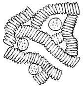
Young folks have a great fancy for "bloody stories;" so I am going to tell you not exactly a "bloody story," but a story about blood, and you need not be alarmed, for before I finish you will find there is nothing in blood to alarm any one, but a great deal that is useful, curious, and beautiful. If you prick your finger with a needle, and squeeze out a drop of blood and place it under the microscope, you will be astonished at what you see (Fig. 14). You can hardly believe that a drop of blood contains so many curiosities. First you observe a whole lot of little reddish-looking bodies, and among these a number of larger transparent bodies, which look like minute splashes of light-colored jelly. It is about these jelly-like bodies I am going to talk with you. If
 | ||
| Fig. 14.—Blood-Cells, colored and colorless. | Figs. 15 and 16.—The Amœba or Blood-Cell changing its Form. | |
you keep your eye on one of them, you see that it continually changes its form, and that it has a slow, crawling kind of motion; and, if you try to make a drawing of it on paper, your picture will never be twice alike (Figs. 15, 16). It puts out something from one side which looks like a foot; then it draws in this foot, and puts out another at the other side, as if trying to find a soft place to walk upon. Sometimes it puts out several of these feet at one time. This little jelly-splash appears to use its feet as we use ours, to walk with, though you see it gets on quite slowly and awkwardly. Its foot is called a pseudopodium, which means false foot. These little bodies have a very suitable name—amœbæ, and the word means changing. This name was given to them, no doubt, because they are constantly changing their form. The amoeba, or blood-cell, is larger than the still protococcus, or mould of the paling, and not quite as large as the moving protococcus, or green water-mould. It is usually about 1/2500 of an inch in breadth. It does not possess the cellulose or woody sac, like the little protococcus houses. It is more like the pear-shaped protococcus boatmen. Its wall is just the hardened outer layer of the jelly or protoplasm. It has no thin space or vacuole, no "eye-spot," no eyelashes or cilia, and when it is quite fresh and new you cannot see any kernel or nucleus. You really can see nothing but an odd-looking lump, with here and there some little grains inside of it. If you give it a drop of weak vinegar or acetic acid, the little grains will disappear, and you can see the kernel in the centre (Figs. 18, 19).
You can find this kernel much easier if you stain the cell with magenta or weak iodine. Heat makes these amœbæ or blood-cells move
 | ||
| Fig. 17. | Figs. 18 and 19.—Cells cleared with Vinegar, so as to show the Nucleus. | |
much quicker, and it is very interesting to watch them make their way among the yellowish-red cells which lie in rows all around them. Sometimes one of the amœbæ will clear a channel for itself right through a thick group of the others. The first time I ever saw them moving in this way, I could not help thinking of the canals in Venice, where the gondoliers steer their gondolas close beside the houses, turning the corners so skillfully as never to strike them. So these little gondoliers of the blood went in and out, threading their way among the reddish-yellow cells, which stood in rows on either side the narrow channels (Fig. 20). There is one great difference, though: in Venice

Fig. 20.—The Amœbæ pushing their Way among the Red Cells.
the gondolier has nothing to do but follow the canals that are made for him, but the amœbæ gondolier has to make his way as he goes. No wonder you open your eyes! It is enough to open any one's eyes to think of the thousands of these odd creatures that go half creeping, half walking through one's veins. You know people sometimes talk about their "blood crawling," but very few people know how the blood crawls.
If you heat the amœbæ they pull in all their little feet and become perfectly white and still, and nothing you can do will ever bring them back to life. These blood-cells that I have described are called "human amœbæ," or the amœbæ of man. Those that are found in the blood of other animals are somewhat different, but there is a kind of amœba, or crawling-cell, which grows on the top of stagnant water, that has a greater difference (Figs. 21, 22). Take a little of the scum that rises on ponds in hot weather, and put it under the microscope. In it you will find these jelly-lumps of a much larger size. They are from 11000 to 1100 of an inch across, and move about by the same kind of queer-looking feet. But the border does not look at all the same. First, on the outside you see a clear, glassy-looking rim; inside of this is a thicker, darker ring filled with little grains—granules.
 | |
| Fig. 21 & 22.—Pond Amœba. | Fig. 23. |
The centre of the cell is quite clear, and contains a thin space or vacuole, like that you saw in the yeast and green mould. The outer border or rim is called the ectosarc, which means outer flesh. The inner is called the endosarc, or inner flesh. Near the clear outer rim or ectosarc you will find the kernel or nucleus—a roundish, solid-looking little body which does not change its form. If you look closely you will see a small round, clear space in this outer rim or ectosarc which has motion, something like the beating of the heart. Indeed, by some it is thought to be the simplest form or beginning of a heart. It is called by a long name, the "contractile vesicle," or "contractile space." All amœbæ do not have this heart, nor do they all have a kernel. This contractile space or heart is very important, because it seems to be doing a work of its own. This is the first time we have found one part of a cell doing something entirely different from another part. The jelly or protoplasm of the yeast and the green mould is "maid-of-all-work." But the amœbæ family seem to be looking up in the world, and are trying to pattern after those establishments that keep a servant for each kind of work.
Inside of some of these large pond amœbæ you will often find the green protococcus-cells, little diatoms, desmids, and all kinds of cells that are smaller than the amœba itself (Fig. 24). These it feeds upon, and, if you have patience to look long enough, you can see how it eats. It has no mouth in particular—the feet seem to taste of what-ever comes in their way, and, if they like it, they grasp it, and poke it in anywhere into the middle of the jelly or protoplasm (Fig. 25).
 | |
| Fig. 24.—Pond Amœba digesting its Food. | Fig. 25.—Amœba eating. |
Here it is digested, and all parts that cannot be used are pushed out again. All that the amœba has to do is to swallow the mass, suck out all the meat, and throw the rest away. There is one thing you must remember about the amœba; it must have its food or protein ready made. It has no power like the yeast or protococcus cell to make it for itself. So these little jelly-lumps we find in the blood and the ponds must be animals. You remember, I told you all vegetables make protein, while all animals eat it up. This little amœba animal gets its full share. He is a perfect little gourmand, taking in every thing that comes in his way. The human amœbae are more fastidious in their taste. They do not swallow their food whole like the wild amœbæ. But those that are found in the blood of the newt or frog are regular little cannibals, and eat up their "colored brethren" whenever they get the chance.
Then, too, the feet or pseudopodia of the savage tribe are thicker and shorter than feet of the civilized kind. You see the toes of your amœba are quite dainty and tapering, like a lady's fingers. It is very curious to watch how a pseudopodium is made, especially of the pond amœba. First there is a little swelling or lifting up of the glassy
 | ||
| Fig. 26.—Wild Amœba. | Fig. 27.—Human Amœba. | Fig. 28.—Amœba making a Foot. |
rim or outer flesh (Fig. 28). As this swelling gets larger, some of the inner flesh flows into it, carrying the little grains, till the swelling is all filled up (Fig. 29).
Then the walking is so funny! The feet do not act as the feet of other animals, carrying the body above them. First, one stumpy foot is put out as far as it can reach, then the body all runs into the foot, and another foot is stuck out from some other part, and away goes the body into this new foot. So it gets on, the feet actually swallowing
 | ||
| Fig. 29.—Grains flowing into Foot. | Fig. 30.—Trying to walk. | Fig. 31.—Stained with Magenta. |
the body! The toad sometimes swallows its old skin, but the amœba is the only animal I know which is "taken in" by its feet! How odd it would be, if, as you walk along, you should suddenly disappear into your boots! If you crush the amœba, you find no trace of a tough sac such as you found in the yeast and protococcus cells. You can see nothing but the kernel or nucleus, and even that soon disappears. If you stain with magenta or iodine, the whole cell becomes colored alike. If there were a tough, woody sac, as in the yeast and mould, it would not be stained. The iodine does not give it a blue color, so there cannot be any starch in the amœbæ. The amœbæ grow like the green-mould cells, by fission, that is, by one or two partitions made through the old cells. You will first see two kernels appear in one of the old cells; then, by close watching, you see a partition going right down between the kernels, separating the old cell into two, with a kernel or
Fig. 32.

nucleus in each. Each new cell follows in the footsteps of its ancestors, crawling, eating, and growing, in the same way. And now I hope you have learned enough about this curious amœbæ family to put your wits to work and give us, some day, a full history of them, and tell us of what use they are, and what they mean by all their motions.
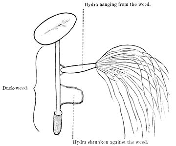
Boys, and girls too, sometimes, love to wade in ditches and ponds in warm weather. When you are thus wading, some time, if you will take up a piece of the duck-weed that grows on the surface of the water, you may see a number of slender green, brown, or orange-colored bodies, about a half-inch long, hanging down from the weed. If you shake or touch them the least bit, they get sulky and shrink

Fig. 33.
all up against the stem, looking like little pouting lumps of jelly. Place your bits of weed in a glass of water, and set the glass in the

Fig. 34. Hydra fishing for Food with its Feelers or Tentacles.
light, but not in the sun, and in a few hours you will find a good many of these little creatures clinging to the side of the glass toward the window. They hold on to the glass by one end, and all around the other end, which is wider, are a number of long threads called tentacles, hanging down gracefully in the water. At first you might think them whiskers, as they grow out around the mouth, but tentacle means a feeler or holder, and with its tentacles you will see how our little friend feels and holds its food, and carries it to his mouth, almost as you use you fingers. These little animals are called hydræ, because if you cut them up each piece will grow again, as did the heads of the old Greek monster. If you look at your hydra under the microscope, you will find all these parts: first, there is the part by which it holds on; it is round and hollow, something like the bottom of a fly's foot, and it changes its size whenever the body of the hydra changes its form. When the hydra is stretched out full length, the foot is smaller than the body; but, when the hydra shrinks up against the glass, it

Fig. 35. Green Hydra.
seems to be all foot. When the body is stretched out, it is round and hollow like a pipe-stem, or more like a very slender funnel, and the opening at the large end surrounded by tentacles or feelers is the mouth.
The hydra's feelers are not all the same length; some of them are prettily colored, and all are filled with wavy knobs or knuckles along the sides (Fig. 35). The bag or body of the hydra is made of two coats. The outer coat is the ectoderm or "outer skin," the other is the endoderm or "inner skin" (Fig. 36). The cells in the outer skin of the green hydra contain those green grains or chlorophyll which give the green color. It is curious to see that the hydra makes its fingers, or tentacles, somewhat as the amoeba makes its feet, or pseudopodiæ (Fig. 36). It pushes out its two coats in the same way, but it never allows its fingers to swallow it as the amœba is swallowed by

Fig. 36.—Hydra pushing out its Fingers.
its feet. When it is disturbed or frightened, it seems to swallow its fingers, or rather puts them all into its mouth, like a sulky child. It is a good deal higher up in the world than the amœba, for you remember that had to eat with its feet. Then, too, the hydra has a more aristocratic walk than the amœba. You can see it plant its foot firmly against the glass, then proudly bow its back and draw the rest of its body up to the foot, in the form of a loop, like the "looping caterpillar" (Fig. 37).
 | ||
| Fig. 37.—Looping Caterpillar. | Fig. 38.—Thread-Cell. | Fig. 39.—Thread-Cell. |
To be sure, it goes backward, but it is a great improvement on the walk of the amœba. It is also an excellent swimmer. You may often see it lift up its foot and dash into the water in search of food. It is one of the funniest things in the world to see the hydra catch its prey. I remember in my old geography a picture of Indians catching wild-horses with lassos. The lasso is a long rope with a loop at the end, which the Indian skillfully throws over the horse's head as he chases it over the prairies. So the hydra throws out his long, rope-like fingers, and lassoes the little animals that swim near it. Sometimes it gets hold of animals so strong that they may tear the fingers or tentacles, and get away again. But the hydra will not be outdone in this way. He has another weapon at hand. Some of the cells in the outer skin are oval, or egg-shaped, and if you look through the cell-walls you see inside what appears to be a long, coiled thread, with two hooks at the bottom (Figs. 38 and 39). These egg-shaped cells are called "thread-cells" and the hydra has many thousands of them in his feelers or tentacles. This thread or spring darts out of its shell whenever the hydra needs it, and sticks itself into the body of the prey like a sharp harpoon (Figs. 40 and 41).
 | |
| Fig. 40—Thread-Cell, with its Thread uncoiled. | Fig. 41.—Thread-Cell, with the Spring turned out like a harpoon. |
If you examine this harpoon closely, you will find that it is only a part of the cell poked in like the finger of a glove turned inward; and when the fingers, or tentacles, seize an animal, these glove-finger cells that cover the tentacles all dart out. Some of them seem to contain a poisonous juice which stupefies or kills the prey in an instant. There is an animal in the sea called the Portuguese man-of-war, which is really a dangerous creature, it has so many of these sharp harpoons. When the prey is stunned or dead, the fingers carry it to the mouth, and it passes down into the long tube or body of the hydra, where it is digested, as though it were in a regular stomach. Along the outside of this funnel-shaped body you may often see little buds, which grow and give off other buds, till the old hydra looks like a branching tree. These buds, no doubt, make you think of something you have seen before—the yeast-babies—yes, these are the baby-hydræ. Soon their fingers begin to grow; then they loosen themselves from the old mother hydra, and begin to "fish for themselves." The next time yon go wading, you must try and capture some of these

Fig. 42.—Old Hydra and Young Ones.
wonderful little creatures, and see if you can find all that I have described without my help. You are now, I trust, opening your eyes to the great world of living things all around you, in which you have lived and played, as I lived and played—blindfolded. And, when once your eyes are really open, wide, there is no telling what wonders they may behold.
- ↑ From "Boys and Girls in Biology," now in the press of D. Appleton & Co., by a pupil of Prof. Huxley. Written upon the basis of his lectures, and illustrated by Miss M. A. J. Macomish.
- ↑ Recent investigations seem to prove that the breathing of plants is similar to that of animals during both day and night; that the breaking up of carbonic acid is digestion, and not respiration. It has its seat in the chlorophyll, and is active in the sun-light; while the respiration, the breathing in of oxygen and the breathing out of carbonic acid, has its seat in the protoplasm, or protein of the cells. (See "Respiration of Plants," by Émile Alglave, Popular Science Monthly for November.)
- ↑ The kind of berry-moulds that grow on old wood and stones, and in the snow, is called still protococcus; that which grows in old water, and moves about by oars, is called moving protococcus.