centres at the anterior end of the body which tend to assume more and more complete control over those lying behind.
Spinal Cord.—A remarkable peculiarity occurs in the sun fishes (Molidae), where the body is greatly shortened and where the spinal cord undergoes a corresponding abbreviation so as to be actually shorter than the brain.
Brain.—It is customary to divide the brain into three main regions, fore-, mid-, and hind-brain, as in the most familiar vertebrates there is frequently seen in the embryo a division of the primitive brain dilatation into three vesicles lying one behind the other. A consideration of the development of the brain in the various main groups of vertebrates shows that these divisions are not of equal importance. In those archaic groups where the egg is not encumbered by the presence of a large mass of yolk it is usual for the brain to show in its early stages a division into two main regions which we may term the primitive fore-brain or cerebrum and the primitive hind-brain or rhombencephalon. Only later does the hinder part of the primitive fore-brain become marked off as mid-brain. In the fully developed brain it is customary to recognize the series of regions indicated below, though the boundaries between these regions are not mathematical lines or surfaces any more than are any other biological boundaries:—
| Rhombencephalon (Hind-brain) | Myelencephalon (Medulla oblongata). | |
| Metencephalon (Cerebellum). | ||
| Cerebrum (Primitive Fore-brain) | Mesencephalon (Mid-brain). | |
| Thalamencephalon (Diencephalon). | ||
| [Hemispheres (Telencephalon).] |


The myelencephalon or medulla oblongata calls for no special remark, except that in the case of Torpedo there is a special upward bulging of its floor on each side of the middle line forming the electric lobe and containing the nucleus of origin of the nerves to the electric organ.

| ||||||||||||||||
| A and B from Wiedersheim, by permission of Gustav Fischer. | ||||||||||||||||
|
Fig. 29.—Brain of Scyllium (A), Salmo (B) and Lepidosiren (C). | ||||||||||||||||
|
The cerebellum occurs in its simplest form in lampreys and Dipnoans (fig. 29, C), where it forms a simple band-like thickening of the anterior end of the roof of the hind-brain. In Selachians it is very large and bulges upwards, forming a conspicuous organ in a dorsal view of the brain (fig. 29, A). In Teleosts (fig. 29, B) the cerebellum is also large. It projects back as a great tongue-like structure over the roof of the fourth ventricle, while in front it dips downwards and projects under the roof of the mid-brain forming a highly characteristic valvula cerebelli. A valvula cerebelli occurs also in ganoids, while in the Crossopterygians a similar extension of the cerebellum projects backwards into the IV. ventricle or cavity of the hind-brain (fig. 30).
The mesencephalon is a conspicuous structure in the fishes from its greatly developed roof (tectum opticum) which receives the end pencils of the optic nerve. Normally it projects upwards as a pair of large optic lobes, but in the Dipnoans (fig. 29, C) the lateral thickening is not sufficiently great to cause obvious lateral swellings in external view.

| ||||||||||||||||||||||||||||||||||||||||||
|
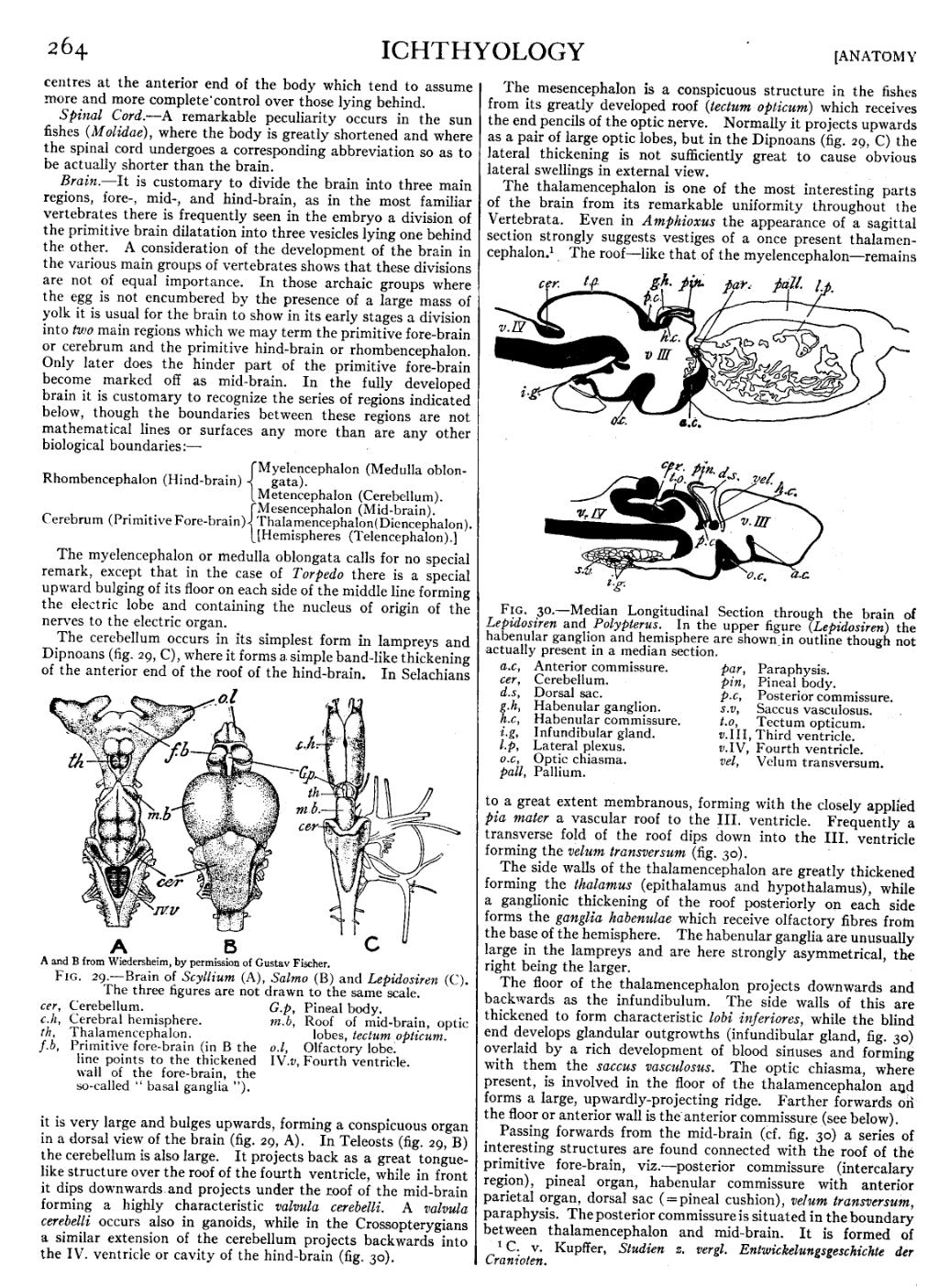
Fig. 30.—Median Longitudinal Section through the brain of Lepidosiren and Polypterus. In the upper figure (Lepidosiren) the habenular ganglion and hemisphere are shown in outline though not actually present in a median section. | ||||||||||||||||||||||||||||||||||||||||||
|
The thalamencephalon is one of the most interesting parts of the brain from its remarkable uniformity throughout the Vertebrata. Even in Amphioxus the appearance of a sagittal section strongly suggests vestiges of a once present thalamencephalon.[1] The roof—like that of the myelencephalon—remains to a great extent membranous, forming with the closely applied pia mater a vascular roof to the III. ventricle. Frequently a transverse fold of the roof dips down into the III. ventricle forming the velum transversum (fig. 30).
The side walls of the thalamencephalon are greatly thickened forming the thalamus (epithalamus and hypothalamus), while a ganglionic thickening of the roof posteriorly on each side forms the ganglia habenulae which receive olfactory fibres from the base of the hemisphere. The habenular ganglia are unusually large in the lampreys and are here strongly asymmetrical, the right being the larger.
The floor of the thalamencephalon projects downwards and backwards as the infundibulum. The side walls of this are thickened to form characteristic lobi inferiores, while the blind end develops glandular outgrowths (infundibular gland, fig. 30) overlaid by a rich development of blood sinuses and forming with them the saccus vasculosus. The optic chiasma, where present, is involved in the floor of the thalamencephalon and forms a large, upwardly-projecting ridge. Farther forwards on the floor or anterior wall is the anterior commissure (see below).
Passing forwards from the mid-brain (cf. fig. 30) a series of interesting structures are found connected with the roof of the primitive fore-brain, viz.—posterior commissure (intercalary region), pineal organ, habenular commissure with anterior parietal organ, dorsal sac ( = pineal cushion), velum transversum, paraphysis. The posterior commissure is situated in the boundary between thalamencephalon and mid-brain. It is formed of
- ↑ C. v. Kupffer, Studien z. vergl. Entwickelungsgeschichte der Cranioten.